Anterior cruciate ligament
| Ligament: Anterior cruciate ligament | ||
|---|---|---|
 |
||
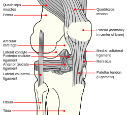
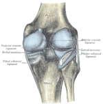
| Diagram of the right knee. (Anterior cruciate ligament labeled at center left.) | ||
| Latin | ligamentum cruciatum anterius | |
| Gray's | subject #93 342 | |
| From | lateral condyle of the femur | |
| To | intercondyloid eminence of the tibia | |
| Dorlands/Elsevier | l_09/12492099 | |
The anterior cruciate ligament (ACL) is one of the four major ligaments of the human knee. In the quadruped stifle (analogous to the knee), based on its anatomical position, it is referred to as the cranial cruciate ligament.[1]
The ACL originates from deep within the notch of the distal femur. Its proximal fibers fan out along the medial wall of the lateral femoral condyle. There are two bundles of the ACL—the anteromedial and the posterolateral, named according to where the bundles insert into the tibial plateau. The ACL attaches in front of the intercondyloid eminence of the tibia, being blended with the anterior horn of the lateral meniscus. These attachments allow it to resist anterior translation of the tibia, in relation to the femur.
Anterior cruciate ligament injury is the most common knee ligament injury, especially in athletes.
Contents |
Additional images
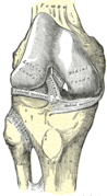 Right knee-joint, from the front, showing interior ligaments. |
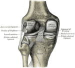 Left knee-joint from behind, showing interior ligaments. |
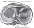 Head of right tibia seen from above, showing menisci and attachments of ligaments. |
 Capsule of right knee-joint (distended). Posterior aspect. |
See also
- Lateral collateral ligament
- Medial collateral ligament
- Posterior cruciate ligament
- Anterior drawer test
- Anterior cruciate ligament reconstruction
References
External links
- SUNY Labs 17:02-0701 - "Major Joints of the Lower Extremity: Knee Joint"
- SUNY Figs 17:07-08 - "Superior view of the tibia."
- SUNY Figs 17:08-03 - "Medial and lateral views of the knee joint and cruciate ligaments."
- lljoints at The Anatomy Lesson by Wesley Norman (Georgetown University) (antkneejointopenflexed)
|
||||||||||||||||||||||||||||||||||||||||||||||||||||||||||||||||||||||||||